Congenital Clubfoot
What is congenital clubfoot?
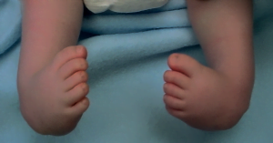
Congenital club foot (also known as talipes equinovarus) is a pathology in which an infant’s foot is turned inward.
It affects approximately one infant in every 1,000 live births, making it one of the more common congenital foot deformities right after hip dysplasia.
Congenital clubfoot can affect both feet (bilateral) with a 35 percent chance and it is more frequent in males than in females.
What causes clubfoot?
The exact cause of clubfoot is not yet known.
There are different theories on its causes but two hypotheses are dominant:
– a defect during embryonic development (genetic factors)
– an incorrect position of the foot during pregnancy in which the uterus presses on the not fully formed skeleton of the child.
Other risk factors may include:
Family history
Other neurological conditions such as Spina Bifida and Cerebral Palsy
Other congenital conditions
Babies with clubfoot may be at increased risk of having Congenital Hip Dysplasia (Developmental Dysplasia of the Hip – DDH).
Diagnosis of clubfoot
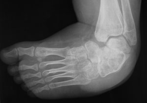
Club foot is present at birth, but with modern prenatal ultrasound examination it is possible to diagnose it around the 22nd week of pregnancy (during morphological ultrasound).
Thanks to an early diagnosis, the orthopedic surgeon can be consulted even before birth and can inform parents about the benign nature of the pathology and plan the course of treatment.
This deformity has very specific characteristics that must be recognized to reach a firm diagnosis.
Congenital clubfoot is a combination of various deformities that affects different joints of the foot.
Clubfoot is usually classified in different groups depending on the deformity and rigidity.
Treatment for clubfoot
Clubfoot is not a painful condition. However, if it is not treated, the foot will remain deformed, and the child will not be able to walk normally.
Most cases of clubfoot are successfully treated with nonsurgical methods that may include a combination of stretching, casting, and bracing.
The most used and successful technique throughout the world is the Ponseti method that consists of two steps:
Manipulation and casting.
The infant’s foot is gently stretched and manipulated into a corrected position and held in place with a cast.
Achilles tendon tenotomy
After the third month of life, if needed, a minor surgery may be required to lengthen Achilles tendon.
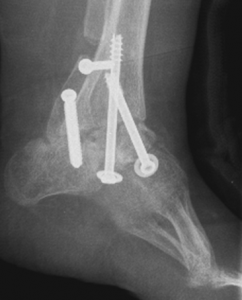
If treatments need to be done at an older age, osteotomy (surgical cut to the bone) may be required. These deformities tends to recur (on average in 10% of the cases).
How Prof. Portinaro treats clubfoot
Prof. Portinaro is one of the most experienced surgeons in the world for foot diseases and treatments.
He has been treating clubfoot for more than 30 years with a non-surgical approach at first, examining more 3,000 patients and performing around 2,500 surgeries with percutaneous tenotomy of the Achilles tendon.
Take a look at this video from Prof Portinaro’s YouTube Channel about Clubfoot in Babies