Legg-Calve-Perthes disease
What is Legg-Calve-Perthes disease
Perthes disease is a rare and temporary childhood condition that affects the hip.
It is a type of osteochondroses, like Kohler disease and Osgood-Schlatter disease.
It occurs because the blood supply to the femoral head (thighbone) is temporarily interrupted. As a result the bone cells begins to die and the area becomes inflamed.
As the condition progresses the dead cells are replaced by new cells that can eventually reshape the head of the thighbone. The mean time for healing is about 50 months.
Legg-Calve-Perthes disease has four main phases that affect the head of the thigh bone:
1 Necrosis
This happen when the blood supply to the femoral head is interrupted. This stage can last for several months.
2 Fragmentation
The body replace dead bone cells with new ones.
This stage can last from 1 to 3 years
3 Reossification
Femoral head develops new and stronger bone cells develop in femoral head. This phase can last for 1 to 2 years.
4 Remodeling and healing
Normal bone cells replace the older bone cells and the femoral head reaches a new rounded final shape
This phase can last a few years.
Legg-Calve-Perthes disease symptoms
Perthes disease usually involves just one hip and one of the earliest signs is limping.
Perthes Disease causes
Perthes disease affects around one out of 12,000 children. Its cause is still unknown but there are some risk factors.
- Age Affected children are mostly between 4 and 10 years old
- Sex Males are 5 times more likely to be affected
- Race White children are more likely to develop it
- Family history Even if in a small number of cases, Perthes disease appeared to run in families.
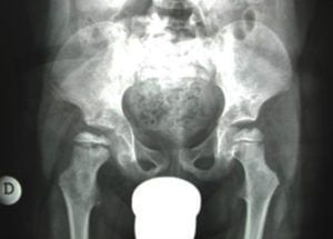
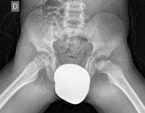
Legg-Calve-Perthes diagnosis
During physical examination the orthopedic surgeon will check the movement of child’s hip and its range of motion.
Legg-Calve-Perthes disease treatment
Treatments depend
- on the severity of the condition
- on the deformity of the femoral head, and its stage of development
- on the child’s age.
The goal of treatment is to keep the head of the thighbone within the acetabulum and to protect its rounded shape as much as possible.
Nonsurgical Treatment may include:
- Observation
- Medications (anti-inflammatory drugs)
- Rest
Surgical Treatment
Surgery may be recommended if:
- nonsurgical treatments proved inadequate
- the child is older than 8 years at the time of diagnosis.
- more than 50% of the femoral head is damaged
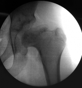
The main goal of surgery is to improve the containment of the femoral head in the acetabulum.
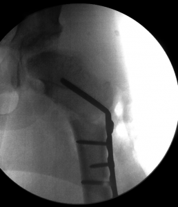
The most common surgical procedure for treating Perthes disease is an osteotomy. The femur bone is cut and repositioned within the acetabulum. Screws and plates are used to keep the femoral head in place. After surgery the patient is kept non weight bearing for 4 weeks. He/She can start early rehabilitation according to pain tolerance.
About Prof. Portinaro’s experience
Prof. Portinaro is one of the most qualified surgeons in the world for hip diseases and treatments, including hip reconstruction.
Prof Portinaro also carried out a research to assess the value of the Magnetic Resonance (MRI) of persistent hip pain in children. MRI was particularly helpful in making early Perthes’ disease diagnosis.